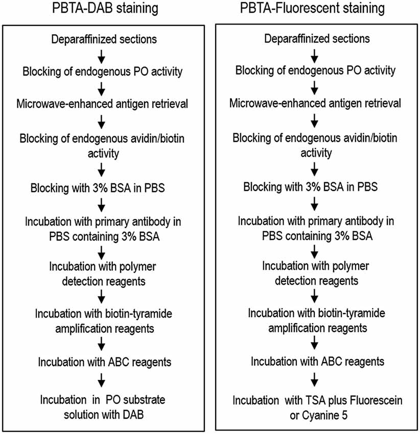
Dab Staining Protocol
DAB Staining Protocol
DAB (3,3'-diaminobenzidine) is a chromogenic substrate that is used in immunohistochemistry and Western blotting to produce a brown precipitate at the site of antigen localization. DAB staining is a simple and sensitive method that can be used to visualize a wide range of antigens, including proteins, carbohydrates, and lipids.

Materials
- Tissue sections or Western blot membranes
- Primary antibody
- Secondary antibody conjugated to horseradish peroxidase (HRP)
- DAB substrate kit
- Hematoxylin (optional)
Procedure
- Deparaffinize and rehydrate tissue sections or soak Western blot membranes in PBS.
- Perform antigen retrieval (if necessary).
- Block tissue sections or membranes with blocking buffer (e.g., 10% BSA in PBS) for 30 minutes at room temperature.
- Incubate with primary antibody for 1-2 hours at room temperature or overnight at 4°C.
- Wash tissue sections or membranes with PBS 3 times for 5 minutes each.
- Incubate with secondary antibody conjugated to HRP for 1 hour at room temperature.
- Wash tissue sections or membranes with PBS 3 times for 5 minutes each.
- Prepare DAB substrate according to the manufacturer's instructions.
- Apply DAB substrate to tissue sections or membranes and incubate for 2-10 minutes, or until desired staining intensity is achieved.
- Rinse tissue sections or membranes with water.
- Counterstain tissue sections with hematoxylin (optional).
- Dehydrate and mount tissue sections or air-dry Western blot membranes.
Tips
- For optimal results, use fresh DAB substrate.
- DAB staining is sensitive to light, so it is important to perform the staining steps in a dark room or under red light.
- Overstaining with DAB can result in a black precipitate. To avoid overstaining, monitor the staining process closely and stop the reaction when the desired staining intensity is achieved.
- DAB staining can be combined with other immunohistochemical stains, such as fluorescent or metal-based stains, to produce multicolor images.
Applications
DAB staining is a versatile technique that can be used to visualize a wide range of antigens in a variety of tissues and cell cultures. DAB staining is commonly used in the following applications:
- Immunohistochemistry: DAB staining is a standard method for detecting proteins and other antigens in tissue sections.
- Western blotting: DAB staining can be used to detect proteins in Western blot membranes.
- ELISA: DAB staining can be used to detect antigens in ELISA plates.
- Flow cytometry: DAB staining can be used to detect antigens on cell surfaces.
DAB staining is a simple and sensitive method that is widely used in research and clinical settings. It is a valuable tool for studying the distribution and expression of antigens in tissues and cells.
