System Biosciences
Mouse Actc Differentiation Reporter (pGreenZeo, Virus)
")
- SKU:
- SR10010VA-1
- Availability:
- Usually Shipped in 5 Working Days
- Size:
- >2 x 10^6 IFUs
- Shipping Temperature:
- Dry Ice
")
")
")
")
Description
Mouse Actc Differentiation Reporter (pGreenZeo, Virus). Cat# SR10010VA. Supplier: SBI System Biosciences


Overview
With SBI’s line of pGreenZeo, pRedZeo, and pRedTK Differentation Reporter Vectors, you can monitor stem cell differentiation in real time. These vectors leverage our reliable lentivector technology and save you time—pre-built differentiation reporters come as ready-to-package lentivector plasmids or ready-to-transduce pre-packaged lentivirus. The Mouse Actc pGreenZeo Differentiation Reporter co-expresses dscGFP (destabilized copGFP, 2-hour half-life) and zeomycin resistance from the mouse Actc promoter, enabling visualization of myogenesis and sorting using GFP fluorescence and selection for the desired cells using zeomycin.
- Create stable myogenesis-reporting cell lines
- Monitor multiple lineages simultaneously
- Track differentiation in live cells in real time
Please note that these vectors only function properly when transduced. Transfection keeps the constitutive RSV promoter intact, leading to nonspecific expression of the reporter genes.
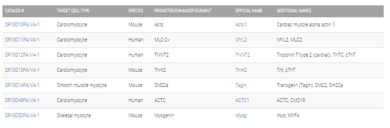
Choose the differentiation reporter that’s right for youMyogenic differentiation reporters
Hematopoietic differentiation reporters

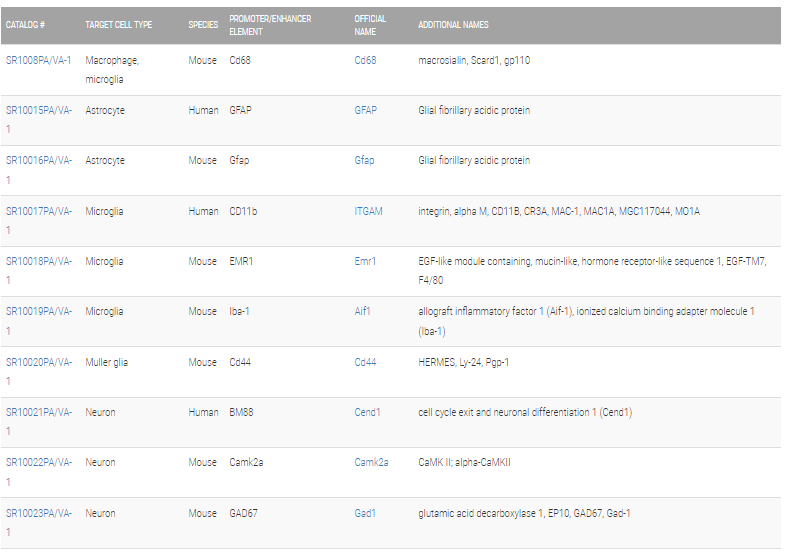
Neural differentiation reporters

Structural differentiation reporters

Signaling differentiation reporters

Supporting Data
See some of our differentiation reporters in action
SBI’s differentiation reporters are used in a number of papers. The data shown below are just one example (from Ravin R, Hoeppner DJ, Munno DM, Carmel L, Sullivan J, Levitt DL, Miller JL, Athaide C, Panchision DM, McKay RD. Potency and fate specification in CNS stem cell populations in vitro. Cell Stem Cell. 2008 Dec 4; 3(6):670-80. PMID: 19041783)

Figure 1. Live imaging of neuronal differentiation. Ravin, et al, used SBI’s Mouse GFAP pGreenFire Differentiation Reporter (Cat.# SR10016VA-1), which drives GFP expression from the glial fibrillary acidic protein promoter, to watch mouse neural stem cells differentiate into a network of mature neurons, oligodendrocytes, and astrocytes over the course of seven days. The periodic “flashes” seen in this video correspond to fluorescent photos taken of the growing cells to identify the GFP signals. The final photo taken after the network formation is shown below the video (color added). Among the network of neurons, only the astrocytes are bright green, demonstrating the specificity of SBI’s mouse GFAP pGreenFire Differentiation Reporter.

Figure 2. Simultaneously track multiple lineages from iPS and progenitor cells.

Figure 3. Additional differentiation reporter data.
Related Products
")
")
Human ACTC Differentiation Reporter (pGreenZeo, Virus)
System Biosciences
")
")
Mouse Actc Differentiation Reporter (pGreenZeo, Plasmid)
System Biosciences
")
")
Mouse Camk2a Differentiation Reporter (pGreenZeo, Virus)
System Biosciences
")
")
Mouse Tnnt2 Differentiation Reporter (pGreenZeo, Virus)
System Biosciences
")
")
Mouse CD8 Differentiation Reporter (pGreenZeo, Virus)
System Biosciences