System Biosciences
Mouse CD68 Differentiation Reporter (pGreenZeo, Virus)
")
- SKU:
- SR1008VA-1
- Availability:
- Usually Shipped in 5 Working Days
- Size:
- >2 x 10^6 IFUs
- Shipping Temperature:
- Dry Ice
")
")
")
")
Description
Mouse CD68 Differentiation Reporter (pGreenZeo, Virus). Cat# SR1008VA. Supplier: SBI System Biosciences


Overview
- Create stable hematopoiesis-reporting cell lines
- Monitor multiple lineages simultaneously
- Track differentiation in live cells in real time


Supporting Data
See some of our differentiation reporters in action
SBI’s differentiation reporters are used in a number of papers. The data shown below are just one example (from Ravin R, Hoeppner DJ, Munno DM, Carmel L, Sullivan J, Levitt DL, Miller JL, Athaide C, Panchision DM, McKay RD. Potency and fate specification in CNS stem cell populations in vitro. Cell Stem Cell. 2008 Dec 4; 3(6):670-80. PMID: 19041783)
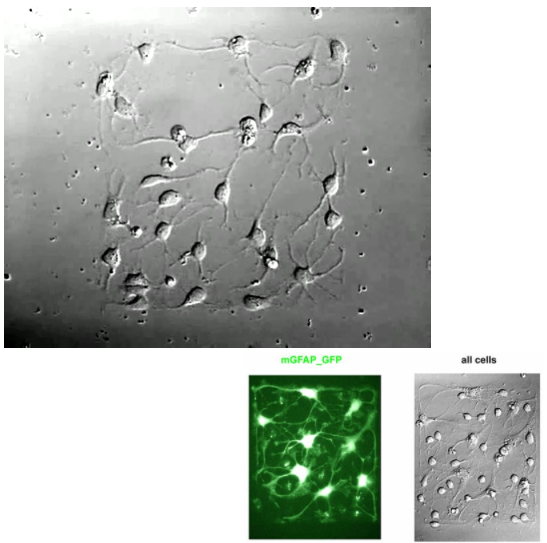
Figure 1. Live imaging of neuronal differentiation. Ravin, et al, used SBI’s Mouse Nestin pGreenFire Differentiation Reporter (Cat.# SR10016VA-1), which drives GFP expression from the glial fibrillary acidic protein promoter, to watch Mouse neural stem cells differentiate into a network of mature neurons, oligodendrocytes, and astrocytes over the course of seven days. The periodic “flashes” seen in this video correspond to fluorescent photos taken of the growing cells to identify the GFP signals. The final photo taken after the network formation is shown below the video (color added). Among the network of neurons, only the astrocytes are bright green, demonstrating the specificity of SBI’s Mouse Nestin pGreenFire Differentiation Reporter.

Figure 2. Simultaneously track multiple lineages from iPS and progenitor cells.
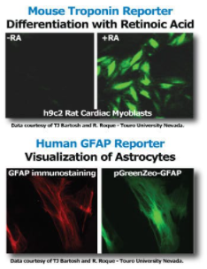
Figure 3. Additional differentiation reporter data.
Related Products
")
")
Mouse CD68 Differentiation Reporter (pGreenZeo, Plasmid)
System Biosciences
")
")
Mouse Camk2a Differentiation Reporter (pGreenZeo, Virus)
System Biosciences
")
")
Mouse Tnnt2 Differentiation Reporter (pGreenZeo, Virus)
System Biosciences
")
")
Mouse CD8 Differentiation Reporter (pGreenZeo, Virus)
System Biosciences
")
")
Mouse GFAP Differentiation Reporter (pGreenZeo, Virus)
System Biosciences