System Biosciences
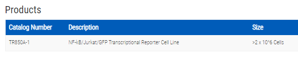
NF-kB/Jurkat/GFP Transcriptional Reporter Cell Line

- SKU:
- TR850A-1
- Availability:
- Usually Shipped in 5 Working Days
- Size:
- >2 x 10^6 cells
- Shipping Temperature:
- Dry Ice


Description
NF-kB/Jurkat/GFP Transcriptional Reporter Cell Line. Cat# TR850A. Supplier: SBI System Biosciences
- Study NF-κB signaling in physiologically-relevant Jurkat cells
- Use for fluorescence microscopy or FACS
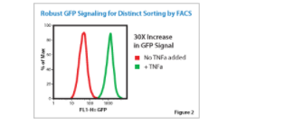
- Take advantage of the cell line’s 30-fold over background NF-κB-dependent GFP expression
- Compatible with a variety of screening methods including small molecule and RNAi
Overview
Monitor NF-κB signaling in Jurkat cells
Speed your studies with this pre-built Jurkat cell line for monitoring NF-κB signaling in real time. We’ve already integrated an expression cassette that includes NF-κB-responsive transcriptional elements upstream of a minimal CMV promoter (mCMV)-GFP cassette. Expression of GFP (up to 30-fold over background) only occurs in the presence of active NF-κB signaling, enabling screening for genetic and/or small molecule inhibitors and activators of the NF-κB signaling pathway.
- Study NF-κB signaling in physiologically-relevant Jurkat cells
- Use for fluorescence microscopy or FACS
- Take advantage of the cell line’s 30-fold over background NF-κB-dependent GFP expression
- Compatible with a variety of screening methods including small molecule and RNAi
Supporting Data
See the NF-κB/Jurkat/GFP Transcriptional Reporter Cell Line in action
Strong GFP fluorescence in response to signaling enables FACS
The NF-κB/Jurkat/GFP provides a strong, dose-dependent response to TNF-α
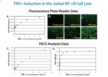
METHODS: NF-B/Jurkat/GFP™ Reporter cells (5×10⁵ cells) were plated at a concentration of 1 million cells/mL into each well of a 24-well plate. TNF-α was added in the amount indicated in the figure. After 24 hours, GFP fluorescence was measured and intensities plotted against TNF-α concentration (A). The fluorescent cells in the original 24-well plate were also photographed on a Zeiss inverted epi-fluorescence microscope (B). Alternatively, 200 μL of cells were fixed with formaldehyde and GFP reporter induction analyzed by flow cytometry, and either the GFP intensities (C) or the percentage of GFP positive cells (D) were plotted against the amount of TNF-α.
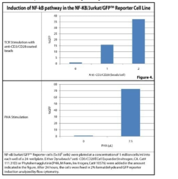
The NF-κB/Jurkat/GFP provides appropriate T cell receptor-mediated signaling
Related Products


NF-kB/293/GFP-Luc Transcriptional Reporter Cell Line
System Biosciences
")
")
MSCV-Human Foxp3-EF1-GFP-T2A-Puro Stable Jurkat cell line (Puro resistant)
System Biosciences
")
")
MSCV-Human RORgt-EF1-GFP-T2A-Puro Stable Jurkat cell line (Puro resistant)
System Biosciences
")
")
MSCV-Mouse Foxp3-EF1-GFP-T2A-Puro Stable Jurkat cell line (Puro resistant)
System Biosciences
")
")
MSCV-Mouse RORgt-EF1-GFP-T2A-Puro Stable Jurkat cell line (Puro resistant)
System Biosciences