System Biosciences
pFC-EF1-MCS-pA-PGK-RFP-T2A-Puro PhiC31 Donor Vector

- SKU:
- FC550A-1
- Availability:
- Usually Shipped in 5 Working Days
- Size:
- 10ug
- Shipping Temperature:
- Blue Ice/ Dry Ice


Description
pFC-EF1-MCS-pA-PGK-RFP-T2A-Puro PhiC31 Donor Vector. Cat# FC550A. Supplier: SBI System Biosciences
- Non-viral transgene delivery
- Single-copy integration
- Preferential integration at active sites
- Delivery of inserts with no size constraints
- Easy generation of cell lines
Products

Overview

- Non-viral transgene delivery
- Single-copy integration
- Preferential integration at active sites
- Delivery of inserts with no size constraints
- Easy generation of cell lines
- Golding MC and Mann MR. A bidirectional promoter architecture enhances lentiviral transgenesis in embryonic and extraembryonic stem cells. Gene Ther. 2011 Aug; 18(8):817-26. PMID: 21390068.
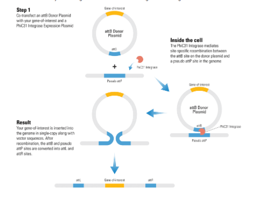
How It Works
One-step transgene delivery with the PhiC31 Integrase System
With the PhiC31 Integrase System, you simply clone your gene-of-interest into the attB Donor Plasmid, and then (Step 1) co-transfect with a PhiC31 Integrase Expression Plasmid. (Inside the cell) The PhiC31 Integrase is transiently expressed, and mediates site-specific recombination between the attB site on the donor plasmid and a pseudo attP site in the genome. (Result) Because pseudo attP sites are typically present in transcriptionally active sites of the genome, your gene-of-interest will likely be integrated into an active region of the genom
Supporting Data
Streamlined gene delivery with the PhiC31 Integrase System

Figure 1. With the PhiC31 Integrase System, the majority of clones that undergo integration events have single insertions.
Southern blot analysis of genomic DNA from positive clones co-transfected with both the PhiC31 Integrase Expression Plasmid (Cat.# FC200PA-1) and the pCOBLW (CAG-Oct4-Sox2-Klf4-Myc-GFP-SV40-Neo) Mouse Reprogramming PhiC31 Donor Plasmid shows the copy number status for 16 different iPSC lines. With both restriction enzymes used, 14 out of 16 lines show single insertions (88%) demonstrating the tight copy number control possible with the PhiC31 Integrase System.

Figure 3. The PhiC31 Integrase System efficiently introduces a puromycin resistance marker and GFP to HEK293 cells.
The dual promoter PhiC31 Donor Vector pFC-PGK-MCS-EF1-GFP-Puro (Cat.# FC551A-1) was co-transfected into HEK293 cells with the PhiC31 Integrase Expression Plasmid (bottom plate), the cells placed under puromycin selection after two days, and cells imaged after another four and eleven days. Comparison of phase versus fluorescence imaging shows that the majority of cells robustly express the GFP reporter.
Related Products


pFC-PGK-MCS-pA-EF1-GFP-T2A-Puro PhiC31 Donor Vector
System Biosciences
 Cloning and Expression Vector")
 Bidirectional Promoter Cloning and Expression Lentivector")
pCDH-EF1-MCS-T2A-RFP (PGK-Puro) Cloning and Expression Vector
System Biosciences


pFC-MCS-pA-SV40-Neo PhiC31 Donor Vector
System Biosciences
 Cloning and Expression Vector")
 Bidirectional Promoter Cloning and Expression Lentivector")
pCDH-EF1-MCS-(PGK-GFP-T2A-Puro) Cloning and Expression Vector
System Biosciences


pFC-CMV-MCS-pA-SV40-Neo PhiC31 Donor Vector
System Biosciences