Pi Staining
Propidium iodide (PI) staining is a technique used to stain DNA and identify dead cells. PI is a fluorescent dye that binds to double-stranded DNA. When PI binds to DNA, it fluoresces red under ultraviolet light.

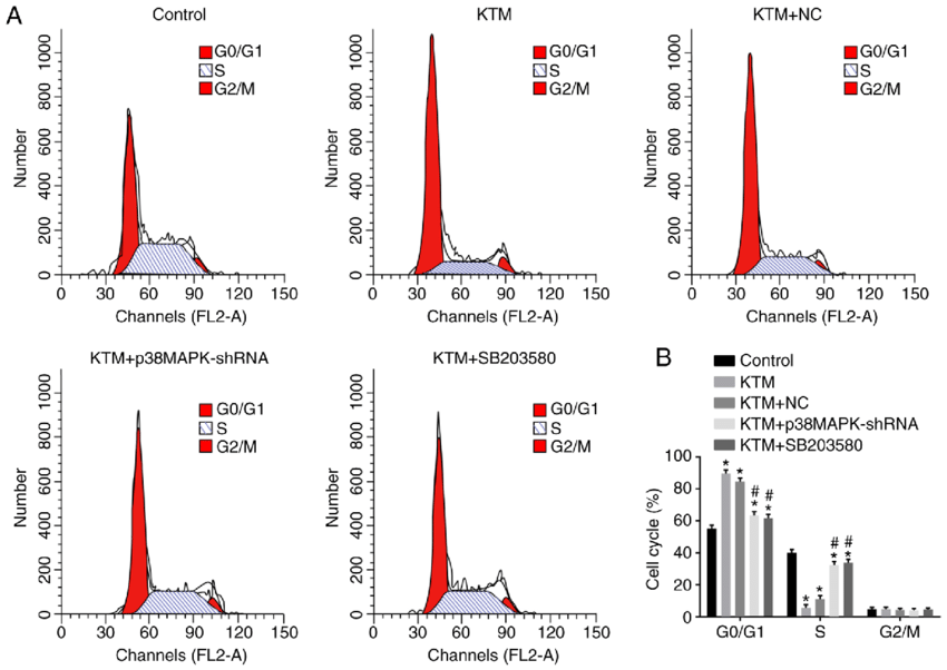
PI staining is commonly used in flow cytometry to measure cell viability. In flow cytometry, cells are stained with PI and then passed through a laser beam. The laser beam excites the PI molecules, which then emit red fluorescence. The amount of red fluorescence emitted by each cell is measured by a detector. Cells with more DNA (i.e., viable cells) will emit more red fluorescence than cells with less DNA (i.e., dead cells).
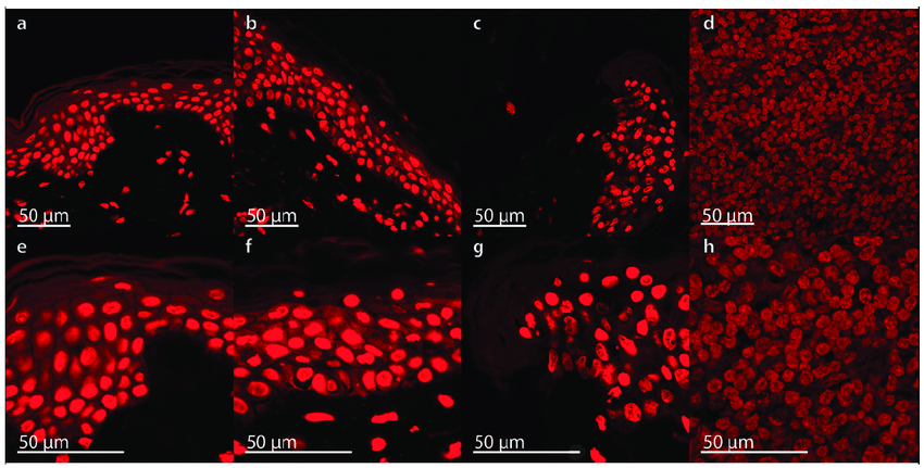
PI staining can also be used in microscopy to visualize DNA and identify dead cells. In microscopy, cells are stained with PI and then viewed under a fluorescence microscope. Viable cells will appear red under the fluorescence microscope, while dead cells will not be visible.
PI staining is a simple and effective method for staining DNA and identifying dead cells. It is used in a variety of research and clinical applications, including:
- Cell biology: PI staining is used to study cell viability, cell cycle progression, and apoptosis.
- Immunology: PI staining is used to identify dead immune cells and to measure the effectiveness of immune responses.
- Cancer biology: PI staining is used to study cancer cell viability, proliferation, and metastasis.
- Clinical diagnostics: PI staining is used to diagnose a variety of diseases, including cancer, HIV/AIDS, and malaria.
PI staining is a valuable tool for biomedical research and clinical diagnostics. It is a simple and effective method for staining DNA and identifying dead cells.
Advantages of PI staining:
- Simple and easy to perform: PI staining is a simple and straightforward technique that can be performed by researchers with basic laboratory skills.
- Reliable and reproducible: PI staining is a reliable and reproducible technique. The results are typically consistent from experiment to experiment.
- Sensitive and specific: PI staining is a sensitive and specific technique for detecting dead cells. It can be used to detect even small numbers of dead cells.
Disadvantages of PI staining:
- Non-specific staining: PI can also bind to other molecules in the cell, such as RNA and proteins. This can lead to non-specific staining and false positives.
- Toxicity: PI is a toxic dye. It can damage cells and tissues if not used properly.
Applications of PI staining:
- Flow cytometry: PI staining is commonly used in flow cytometry to measure cell viability. This is done by passing cells stained with PI through a laser beam and measuring the amount of red fluorescence emitted by each cell. Cells with more DNA (i.e., viable cells) will emit more red fluorescence than cells with less DNA (i.e., dead cells).

- Microscopy: PI staining can also be used in microscopy to visualize DNA and identify dead cells. Cells stained with PI are viewed under a fluorescence microscope. Viable cells will appear red under the fluorescence microscope, while dead cells will not be visible.
- Cell culture: PI staining can be used to study cell viability in cell culture. Cells are stained with PI and then viewed under a microscope to determine the percentage of dead cells.
- Tissue sections: PI staining can also be used to study cell viability in tissue sections. Tissue sections are stained with PI and then viewed under a microscope to determine the percentage of dead cells.
Safety precautions:
- PI is a toxic dye and should be handled with care.
- Wear gloves and eye protection when handling PI.
- Avoid contact with skin and mucous membranes.
- If PI comes into contact with skin or mucous membranes, wash the area immediately with soap and water.
- If PI is ingested, seek medical attention immediately.
Overall, PI staining is a valuable tool for biomedical research and clinical diagnostics. It is a simple and effective method for staining DNA and identifying dead cells. However, it is important to be aware of the potential drawbacks of PI staining, such as non-specific staining and toxicity.
