System Biosciences
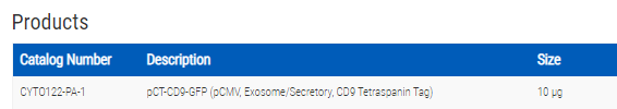
pCT-CD9-GFP (pCMV, Exosome/Secretory, CD9 Tetraspanin Tag)
")
- SKU:
- CYTO122-PA
- Availability:
- Usually Shipped in 5 Working Days
- Shipping Temperature:
- Blue Ice/ Dry Ice
")

")

Description
pCT-CD9-GFP (pCMV, Exosome/Secretory, CD9 Tetraspanin Tag). Cat# CYTO122-PA. Supplier: SBI System Biosciences
- Stable lentivector-based system
- Ideal for co-localization studies
- Monitor exosome dynamics and functional studies in real time
- Label exosomes from primary cells, tumor cells, stem cells, and more
Overview
- Stable lentivector-based system
- Ideal for co-localization studies
- Monitor exosome dynamics and functional studies in real time
- Label exosomes from primary cells, tumor cells, stem cells, and more

Supporting Data
See some of our exosome Cyto-Tracers in action
The following figure and videos are from:
Garcia NA, et al. Glucose Starvation in Cardiomyocytes Enhances Exosome Secretion and Promotes Angiogenesis in Endothelial Cells. PLoS ONE. 2015. 10(9). PMCID: PMC4578916.
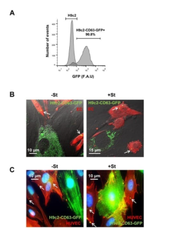
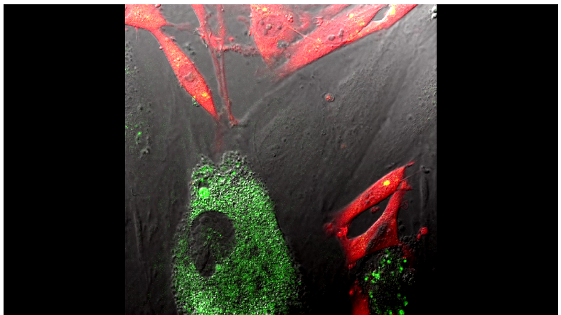
Figure 4 from Garcia, et al. Exosome transfer from CMs to ECs. (A) H9c2 transfected with pCT-CD63-GFP. FACS analysis of 90% GFP positive cells. (B) Representative images from confocal time-lapse microscopy of mouse ECs (ACTB-DsRed) (EC; red) co-cultured with H9C2-CD63-GFP cells (green), previously cultured for 24 h in +/-St medium. Exosome transfer from H9C2 CMs to EC can be observed (S1 and S2 Movies, below). White arrows show CD63-GFP structures inside ECs (C) Representative immunostaining of H9C2-CD63-GFP and HUVEC co-cultures; anti-GFP (green) and anti-CD31 (red). The images illustrate GFP fluorescence from CD63-GFP exosomes in CD31-positive cells (red) after 24 h incubation in +/-St medium. White arrows show CD63-GFP structures inside ECs.
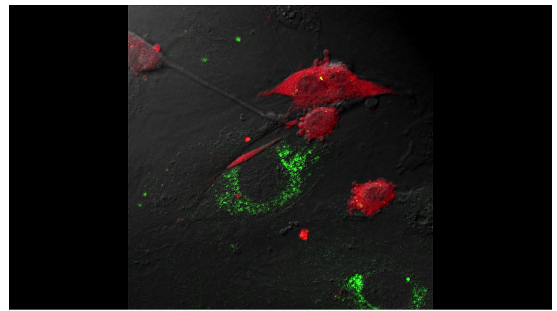
Video S1 from Garcia, et al. Exosome transfer from H9C2-CD63-GFP (green) to endothelial DsRed cells (red) under +St conditions.
Video S2 from Garcia, et al. Exosome transfer from H9C2-CD63-GFP (green) to endothelial DsRed cells (red) under -St conditions.

Labeled exosomes. (Top panels) CD63-GFP Cyto-Tracers transfected into a human fibrosarcoma cell line. (Bottom panels) CD9-GFP and CD9-RFP Cyto-Tracers co-transfected into HEK293 cells.
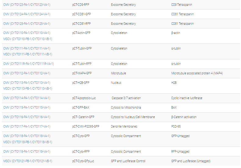
Related Products
")

pCT-CD9-RFP (pCMV, Exosome/Secretory, CD9 Tetraspanin Tag)
System Biosciences
")
")
pCT-CD63-GFP (pCMV, Exosome/Secretory, CD63 Tetraspanin Tag)
System Biosciences
")

pCT-CD81-GFP (pCMV, Exosome/Secretory, CD81 Tetraspanin Tag)
System Biosciences
")

pCT-CD63-RFP (pCMV, Exosome/Secretory, CD63 Tetraspanin Tag)
System Biosciences
")

pCT-CD81-RFP (pCMV, Exosome/Secretory, CD81 Tetraspanin Tag)
System Biosciences