System Biosciences
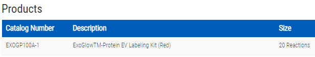
ExoGlow-Protein EV Labeling Kit (Red)
")
- SKU:
- EXOGP100A-1
- Availability:
- Usually Shipped in 5 Working Days
- Size:
- 20 reactions
- Shipping Temperature:
- Blue Ice
")
")
")
")
Description
ExoGlow-Protein EV Labeling Kit (Red) . Cat# EXOGP100A. Supplier: SBI System Biosciences
Overview
- Specific—carefully developed to generate a robust signal specific for internal EV proteins, leading to very low levels of background
- Compatible—delivers robust performance on EVs isolated using all methods tested—including ExoQuick®, ultracentrifugation, and column-based workflows
- Easy-to-use—labeling protocol is quick and straightforward
- Powerful—can be used with as little as 200 µg of EVs
Emission: 588 nm
Laser line: 561 nm
Available next generation ExoGlow EV imaging reagents
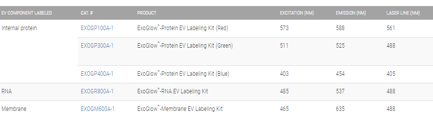
Imaging EVs with a new level of clarity
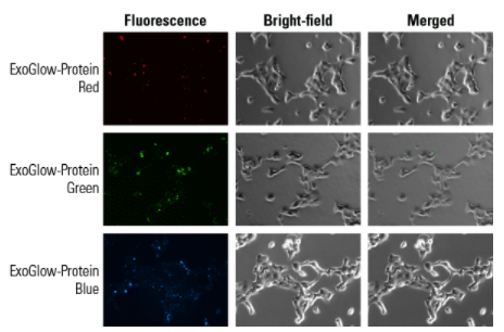
Figure 1. ExoGlow-Protein enables clear visualization of labeled EVs being internalized by target cells. We labeled HEK293T EVs with ExoGlow-Protein and followed uptake by HEK293T cells. The punctate fluorescence signal shows internalization of labeled EVs by the target cells.
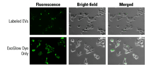
Figure 2. Punctate staining pattern is EV-specific. The punctate staining pattern seen in Figure 1 and Panel A is due to ExoGlow-Protein-labeled EVs. When ExoGlow-Protein is added directly to target HEK203T cells (Panel B), only diffuse, non-specific staining is seen.
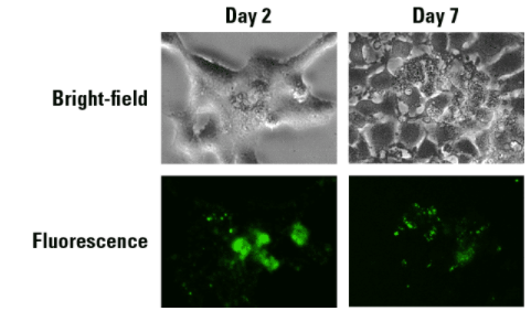
Figure 3. ExoGlow-Protein signal is stable for up to at least 7 days after administration of labeled EVs to cells.
Related Products
")
")
ExoGlow-Protein EV Labeling Kit (Green)
System Biosciences
")
")
ExoGlow-Protein EV Labeling Kit (Blue)
System Biosciences


ExoGlow-RNA EV Labeling Kit
System Biosciences


ExoGlow-Membrane EV Labeling Kit
System Biosciences
")
")
ExoGlow-Vivo EV Labeling Kit (Near IR)
System Biosciences